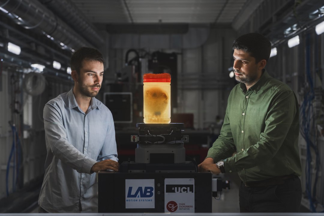
Preparing a human heart for HiP-CT imaging at the ESRF with Hector Dejea. Image: ESRF / Stef Candé.
Revealing human anatomy across scales
I am Dr Joseph Brunet, a Research Fellow at University College London, based at the European Synchrotron Radiation Facility in Grenoble. I specialise in non-destructive, multiscale imaging of intact human organs using Hierarchical Phase-Contrast Tomography (HiP-CT).
My research sits at the intersection of biomedical imaging, cardiovascular science, biomechanics and 3D image analysis. I am particularly interested in how the architecture of the human heart—from myocardial organisation and vascular networks to the conduction system—relates to function and disease.
Research
I lead UCL’s HiP-CT research activities at the ESRF and have coordinated more than 45 experiments on intact human organs. My work spans the full imaging workflow: sample preparation, multiscale synchrotron acquisition, reconstruction and quality control of terabyte-scale volumes, and quantitative analysis.
To extract meaningful measurements from these datasets, I develop methods for segmentation, vascular-network characterisation, structure-tensor analysis, tractography and digital volume correlation. I created Cardiotensor, an open-source Python library for quantifying myocardial organisation in large 3D cardiac images, published in the Journal of Open Source Software in 2026.
My current programme aims to connect micro-anatomy with clinical imaging, biomechanics and molecular information.
Expertise
- Biomedical and synchrotron X-ray imaging
- HiP-CT experimental design and large-volume reconstruction
- Cardiac microstructure, vascular networks and conduction anatomy
- Quantitative 3D image analysis, tractography and segmentation
- Biomechanical testing and computational modelling
- Open research infrastructure and international scientific coordination