Joseph Brunet
  • About
  • Research
  • Publications

Research

Welcome to my research section. Here you’ll find summaries of key areas I work on:

Human Organ Atlas

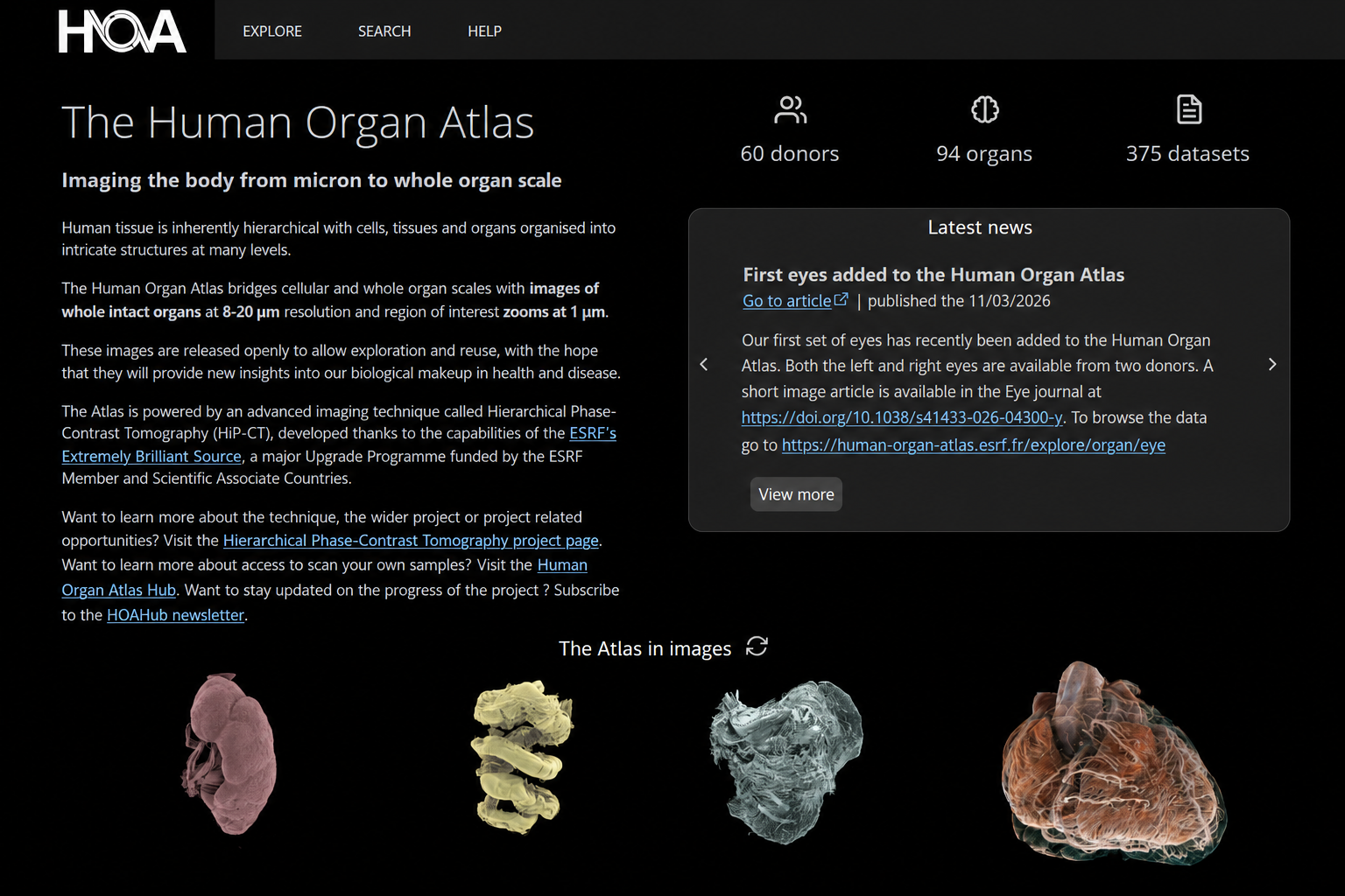
Multiscale 3D Imaging HiP-CT Open Data A “Google Earth” for human anatomy The Human Organ Atlas is an open, interactive resource for exploring intact human organs in three dimensions. It brings together some of the most detailed whole-organ images yet produced, allowing researchers, clinicians, students and the public to move seamlessly from organ-scale anatomy to microscopic structures. The Atlas includes multiscale datasets from organs such as the heart, brain, lungs, kidneys and liver. Because the data can be explored directly in a web browser, users can investigate complex anatomy without specialist visualisation software. ...
Cardiotensor

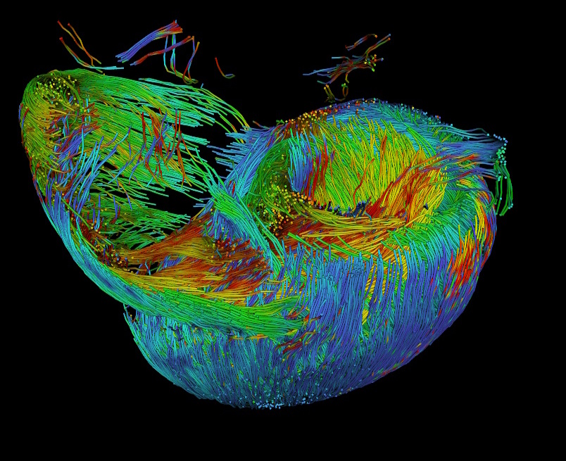
Python Library Tractography Myocyte Orientation Cardiotensor ( ) is an open-source Python package developed to quantify 3D cardiomyocyte orientation and reconstruct continuous tractography of myoaggregates in volumetric cardiac imaging datasets. The library is designed for modern high-resolution modalities such as synchrotron tomography (HiP-CT), micro-CT, and 3D optical imaging. It supports datasets up to teravoxel scale through chunk-based and parallelized processing pipelines. Cardiotensor workflow: orientation computation with structure tensor, helical angle (HA) and intrusion angle (IA) mapping, and streamline-based tractography ...

The Heart Atlas

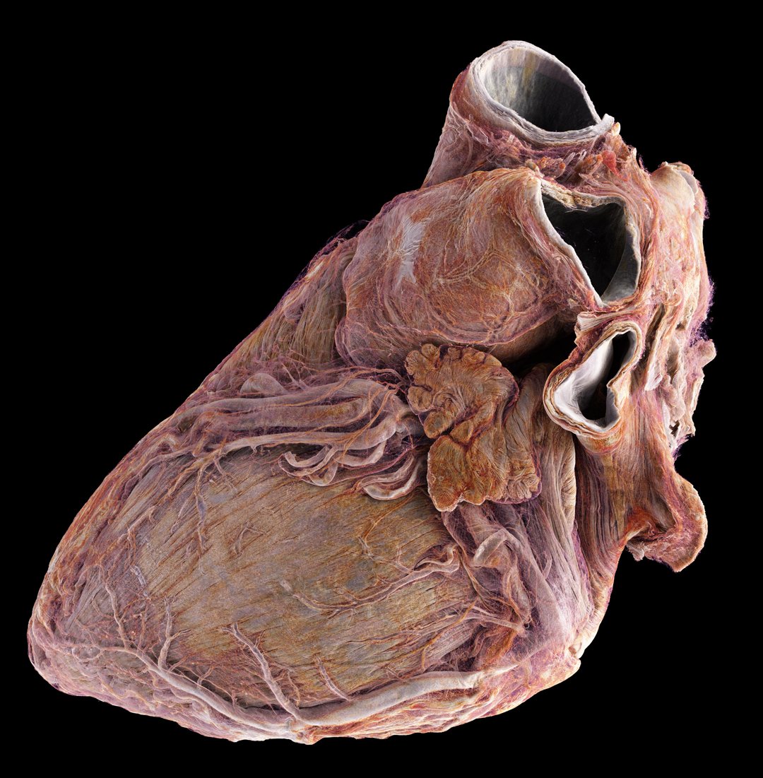
Cardiac Imaging HiP-CT Synchrotron X-ray Tomography During my postdoc at the European Synchrotron Radiation Facility (ESRF), I used Hierarchical Phase-Contrast Tomography (HiP-CT) to depict the macro- to microanatomy of structurally normal and abnormal adult human hearts ex vivo. Joseph Brunet (left) and Hector Dejea (right) positioning a heart during an experiment (Image credit: ESRF / Stef Candé) HiP-CT allows for high-spatial-resolution, three-dimensional imaging of the heart, revealing histologic-level detail without the need for invasive sectioning or exogenous contrast agents. The study involved imaging two hearts: one from a healthy 63-year-old male and another from an 87-year-old female with a history of cardiovascular diseases. ...

Aortic Dissection

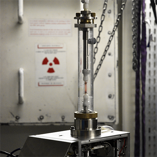
Aortic Dissection Tension-Inflation Synchrotron X-ray Tomography During my PhD at École des Mines de Saint-Étienne, I worked on vascular biomechanics as part of the AArteMIS project. My research focused on identifying and quantifying the mechanisms behind vascular failure, particularly aortic dissection, to improve diagnostic and therapeutic strategies. Suture of a rabbit aortic sample on the tension-inflation device connectors To this end, I performed in situ experiments combining mechanical testing and imaging techniques. I generated in vitro dissections in porcine and rabbit aortas and visualized them using X-ray micro-tomography, both conventional and synchrotron-based. ...

| | |