During my PhD at École des Mines de Saint-Étienne, I worked on vascular biomechanics as part of the AArteMIS project. My research focused on identifying and quantifying the mechanisms behind vascular failure, particularly aortic dissection, to improve diagnostic and therapeutic strategies.

Suture of a rabbit aortic sample on the tension-inflation device connectors
To this end, I performed in situ experiments combining mechanical testing and imaging techniques. I generated in vitro dissections in porcine and rabbit aortas and visualized them using X-ray micro-tomography, both conventional and synchrotron-based.

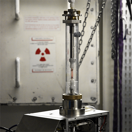
Our tension-inflation device at the ESRF synchrotron
This approach enabled 3D observation of the delamination during dissection propagation through the vessel wall.

2D X-ray radiographs of dissection propagation in real time
Using tensile tests combined with inverse methods, I quantified both the elastic and failure properties of the dissected aortas.

Tensile tests on a porcine aorta in circumferential (left) and axial (right) directions
Finally, I used finite element modeling to simulate rupture and study the fracture modes using XFEM (eXtended Finite Element Method).

Numerical simulation of an aortic dissection using XFEM
© 2022 – Joseph Brunet