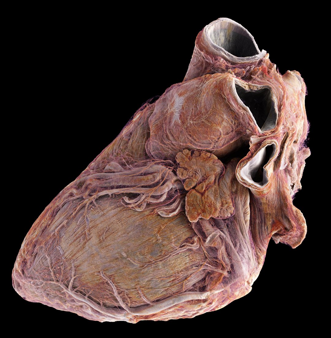
During my postdoc at the European Synchrotron Radiation Facility (ESRF), I used Hierarchical Phase-Contrast Tomography (HiP-CT) to depict the macro- to microanatomy of structurally normal and abnormal adult human hearts ex vivo.
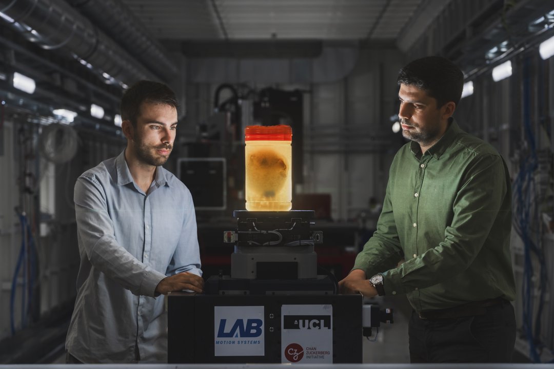
Joseph Brunet (left) and Hector Dejea (right) positioning a heart during an experiment (Image credit: ESRF / Stef Candé)
HiP-CT allows for high-spatial-resolution, three-dimensional imaging of the heart, revealing histologic-level detail without the need for invasive sectioning or exogenous contrast agents. The study involved imaging two hearts: one from a healthy 63-year-old male and another from an 87-year-old female with a history of cardiovascular diseases.
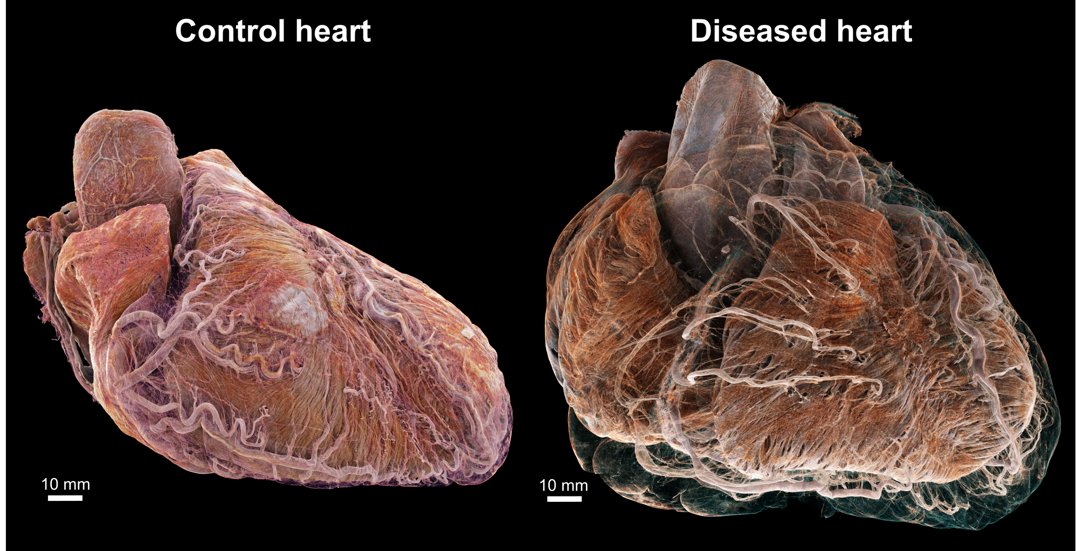
3D cinematic renderings of control and diseased hearts
The imaging revealed intricate details of the myocardium, valves, coronary arteries, and conduction system, with resolutions from 20 µm/voxel down to 2 µm/voxel, allowing to observe some cells such as myocytes.
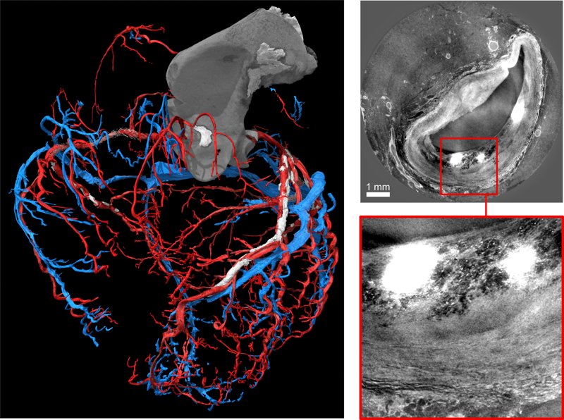
Segmentation of coronary arteries and veins in the diseased heart
HiP-CT also enabled the visualization of pathologic features such as fatty infiltration, vascular supply disruptions, and structural changes due to diseases like myocardial infarction and atrial fibrillation.
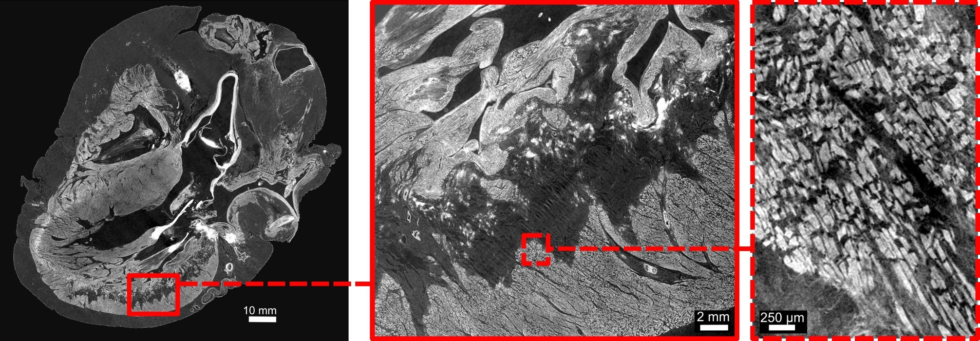
HiP-CT scans of ventricular myocardium in the diseased heart
This study demonstrates the potential of HiP-CT for detailed, non-destructive imaging of the heart, which can greatly enhance our understanding of cardiac anatomy and disease, paving the way for better diagnostic and therapeutic strategies.
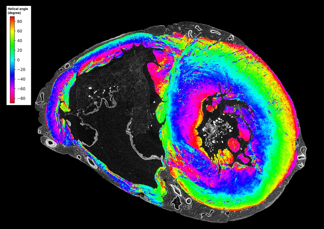
Orientation of myocyte aggregates in the control heart
📄 For full details, see the published paper in Radiology (2024).
© 2024 – Joseph Brunet