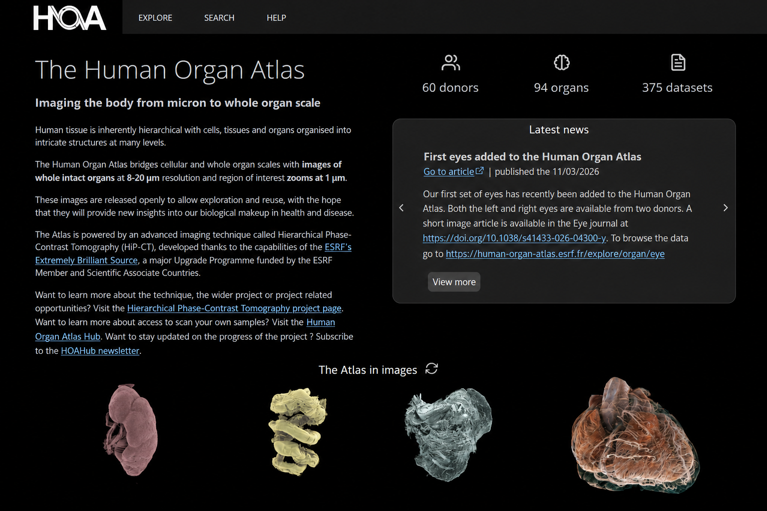
A “Google Earth” for human anatomy
The Human Organ Atlas is an open, interactive resource for exploring intact human organs in three dimensions. It brings together some of the most detailed whole-organ images yet produced, allowing researchers, clinicians, students and the public to move seamlessly from organ-scale anatomy to microscopic structures.
The Atlas includes multiscale datasets from organs such as the heart, brain, lungs, kidneys and liver. Because the data can be explored directly in a web browser, users can investigate complex anatomy without specialist visualisation software.
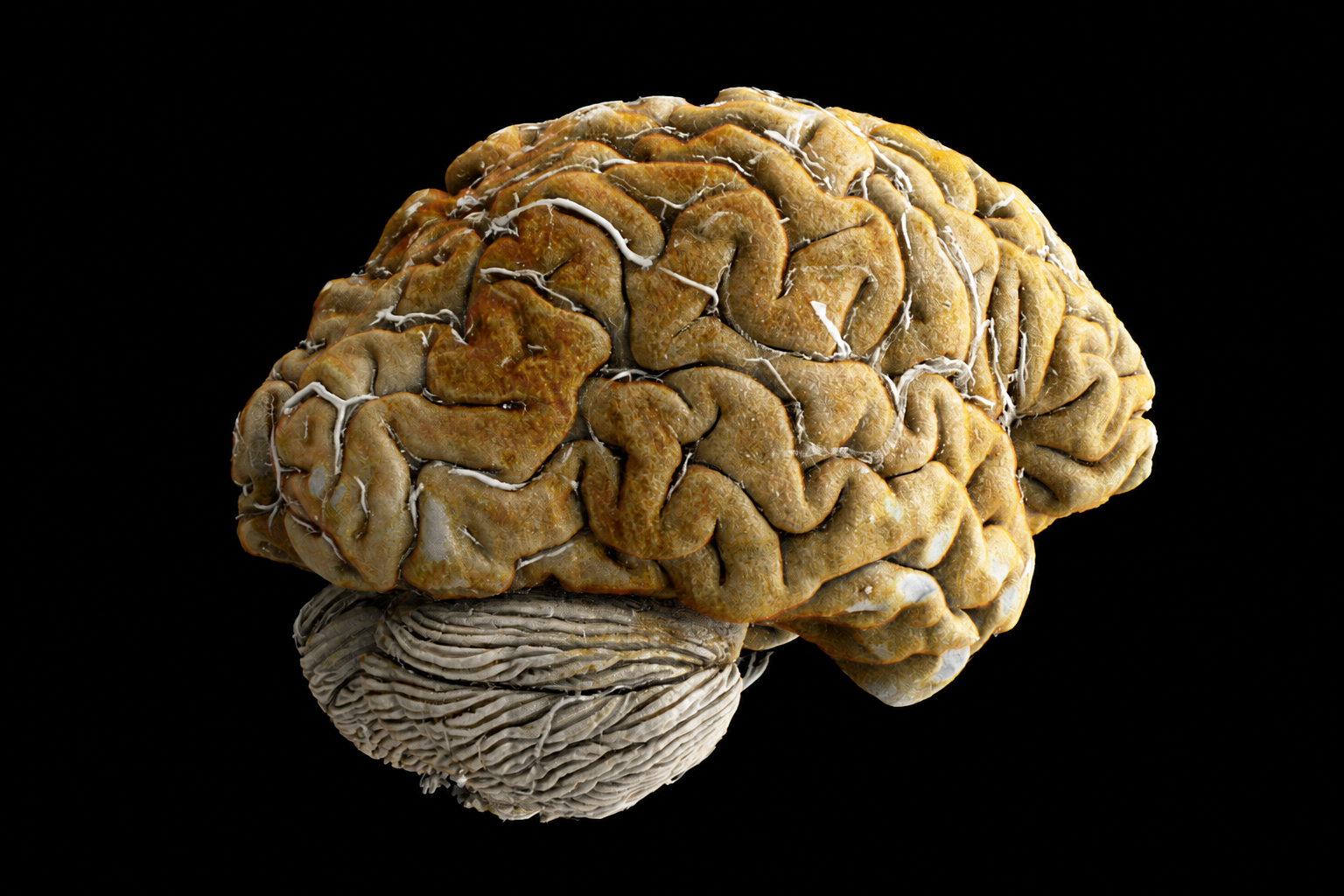
A whole-organ brain visual inspired by Human Organ Atlas imagery, illustrating anatomy preserved in three dimensions.
Imaging across scales with HiP-CT
The Atlas is powered by Hierarchical Phase-Contrast Tomography (HiP-CT) at the European Synchrotron Radiation Facility. HiP-CT overcomes the usual trade-off between field of view and resolution: an entire organ is first imaged at anatomical scale, then selected regions can be scanned at progressively higher resolution without physically sectioning the sample.
This hierarchy connects information normally separated across clinical CT, microscopy and histology. Structures can be followed continuously through the organ, preserving their three-dimensional context and revealing how local changes relate to global anatomy and disease.
From images to an open research resource
My work contributes to the experimental and quantitative foundations of the Atlas: preparing and imaging intact organs at the ESRF, coordinating HiP-CT experiments, reconstructing large multiscale volumes, and developing methods that turn these images into measurable anatomical information.
A major focus is the human heart, including myocardial organisation, vascular networks and the cardiac conduction system. The same open infrastructure also enables research across neurological, respiratory, renal and other organ systems, and provides reference data for validating clinical imaging and computational methods.
Open science and reuse
The Human Organ Atlas makes processed imaging data and associated metadata openly available for scientific reuse. These datasets support anatomical discovery, education, segmentation challenges, machine-learning development and comparison with modalities such as MRI, clinical CT and histology.
The project is described in “The Human Organ Atlas,” published in Science Advances in 2026.